




Quantum dots enable the quantification of misfolded proteins within cells to detect pathological signs of ALS
26.05.2026
 |
|
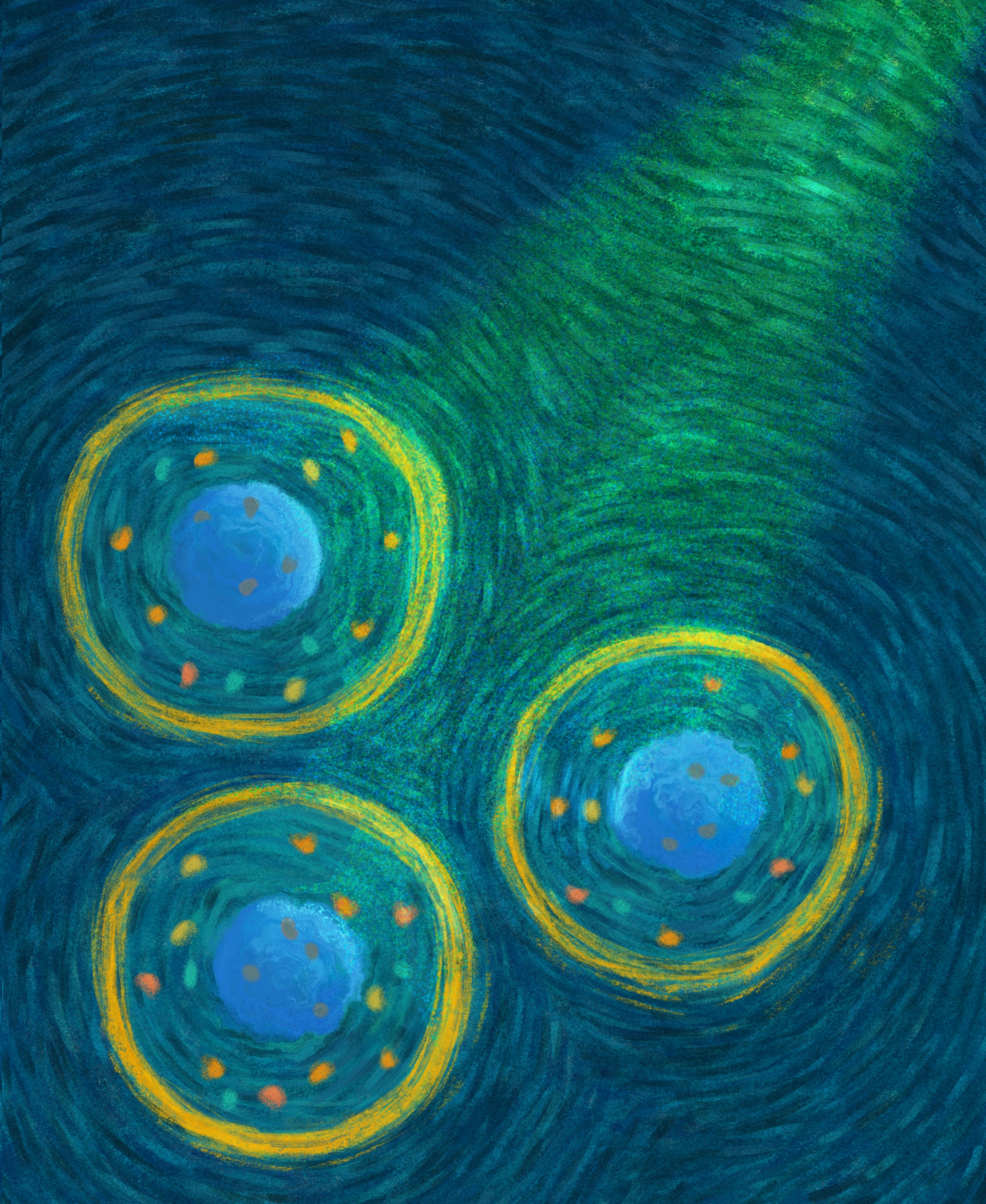
Image: Patricia Bondía / Scientific Videogames. |
- A new strategy makes it possible to identify mislocalized proteins in the cells of patients with amyotrophic lateral sclerosis (ALS).
- The technique combines nanotechnology and flow cytometry to rapidly detect biomarkers, which is essential in clinical practice.
| Tweet |  |
Press release download |
Madrid, 26th May, 2026. The change in the location of certain proteins within cells is a hallmark of amyotrophic lateral sclerosis (ALS). Under normal conditions, certain proteins perform their functions within the cell nucleus; however, in patients with ALS, some of these proteins accumulate in the cytoplasm, while their numbers in the cell nucleus decrease. Fluorescent markers can be used to quantify these proteins, as they illuminate the proteins regardless of their location within the cell. To detect this relocalization, fluorescence microscopy is required, which allows for the precise visualization of these proteins’ locations in individual cells. While this technique is reliable for analyzing individual cells, it is not the most suitable method for analyzing large numbers of cells from different patients—a critical requirement in clinical practice.
A team of researchers at IMDEA Nanociencia, led by Valle Palomo, has developed a strategy that enables the rapid detection of this protein mislocalization using standard clinical equipment, as a potential biomarker for amyotrophic lateral sclerosis (ALS). The strategy is based on quantum dots, nanoparticles that can emit light in different colors and whose signal is highly stable. By using quantum dots linked to antibodies or biomolecules that recognize specific proteins, the team was able to detect and quantify various proteins in the cells under investigation—preserved lymphocytes derived from ALS patients. The key difference between this method and the use of traditional fluorescent markers is that the quantum dots do not penetrate the cell nucleus of the lymphocytes, labeling only the proteins present in the cytoplasm. By taking advantage of this phenomenon, the researchers were able to quickly and efficiently measure the relocalization of the TDP-43 protein outside the cell nucleus. The relocalization of this protein is one of the most significant markers of ALS, and they were also able to simultaneously analyze three other proteins using four different colors of quantum dots. The results show that patients’ cells have significantly higher levels of TDP-43 outside the nucleus than those of healthy individuals.
In addition to analyzing cultured cells, the team validated the technique using mouse brain tissue in collaboration with the Cajal Center for Neuroscience (CSIC). The experiments confirmed that the quantum dots remain outside the cell nucleus and suggest that the strategy could be used to rapidly identify pathological protein aggregates relevant to the study of neurodegenerative diseases.
The ability to measure proteins in only a specific part of the cell represents a key breakthrough and has direct applications in the most widely used technique in hospitals for analyzing blood cells: flow cytometry. Unlike microscopy, flow cytometry does not visualize the cell itself, but only the intensity of the fluorescence it emits; therefore, it is not possible to determine whether the signal originates from the nucleus or the cytoplasm. Researcher Valle Palomo’s quantum dot-based strategy is key in this regard. Since these nanoparticles do not penetrate the nucleus of lymphocytes, the fluorescence detected by flow cytometry reflects only the amount of protein present in the cytoplasm. Thanks to the CaixaImpulse project, funded by the ‘La Caixa’ Foundation, and in collaboration with the Research Institute of the Hospital Clínico San Carlos, the researchers have analyzed cells from ALS patients, obtained through a conventional blood test.
The development of this technique has not been without its challenges, as the amount of protein and the permeability of cells to quantum dots vary from person to person. To address this, the team designed a normalization method that allows for reliable comparison of samples. So far, they have analyzed cells from five patients and plan to expand the study to validate it clinically. The results are demonstrating the technique’s clear potential as a rapid screening tool.
According to Valle Palomo, the study’s lead author, this approach opens up new possibilities for studying the biology of the disease: “Quantum dots act as an indicator of protein delocalization, allowing us to detect proteins outside the nucleus using flow cytometry, a very fast, high-throughput technique that normally does not provide spatial information.” In comparative experiments, when conventional fluorophores were used, no differences were observed between healthy cells and those from patients, because all proteins were labeled without distinguishing their location. With quantum dots, however, the signal comes only from the cytoplasm, and the differences become evident.
The results have been published in the journal ACS Sensors and are part of a project that combines nanotechnology, biomedicine, and diagnostics. The work is the result of a collaboration between researchers at IMDEA Nanociencia, the Health Research Institute of the Hospital Clínico San Carlos, the Cajal-CSIC Neuroscience Center, and the “Margarita Salas” Biological Research Center -CSIC and has been partially funded by the State Research Agency, the ‘La Caixa’ Foundation, and the Severo Ochoa Excellence accreditation awarded to IMDEA Nanociencia.
Keywords: Quantum Dots, amyotrophic lateral sclerosis, TDP-43, protein mislocalization, multiplexed protein detection.
Glossary:
- Amyotrophic lateral sclerosis (ALS): A disease of the nervous system that affects the neurons in the brain and spinal cord. It occurs when cells in the nervous system progressively degenerate, leading to muscle paralysis. The exact cause is unknown, and in a small number of cases (5%), it is hereditary.
- Flow cytometry: an advanced laboratory technique that analyzes, quantifies, and classifies individual cells or particles (such as blood, bone marrow, or tumor cells) in liquid suspension. It can analyze thousands of cells per second and is essential for diagnosing leukemias, lymphomas, and other studies of cell viability.
Reference:

Contact:
Valle Palomo, PhD
This email address is being protected from spambots. You need JavaScript enabled to view it.
Biosensors in Neuroscience Group
https://www.nanociencia.imdea.org/biosensorsinneuroscience/home
IMDEA Nanociencia Dissemination and Communication Office
divulgacion.nanociencia [at]imdea.org


Source: IMDEA Nanociencia.
IMDEA Nanociencia Institute is a young interdisciplinary research Centre in Madrid (Spain) dedicated to the exploration of nanoscience and the development of applications of nanotechnology in connection with innovative industries.
Related information:
Scientists analyze Amyotrophic Lateral Sclerosis patient cells to improve strategy testing https://www.nanociencia.imdea.org/biosensorsinneuroscience/news/item/towards-personalized-treatments-for-amyotrophic-lateral-sclerosis-scientists-analyze-patient-cells-to-improve-strategy-testing?category_id=1479
Valle Palomo receives a CaixaImpulse Health Innovation grant from the “la Caixa” Foundation https://www.nanociencia.imdea.org/biosensorsinneuroscience/news/item/valle-palomo-receives-a-caixaimpulse-health-innovation-grant-from-the-la-caixa-foundation?category_id=1479